Monday, January 28, 2019

Enteropathogenic E.coli infection:


·         Most strains of E.coli are common members of the enteric microflora in the human colon and are not pathogenic. Few strains are potential foodborne pathogens.
·         They produce potent enterotoxins and may cause life-threatening diarrheal disease and urinary tract infections.
Six virulence groups of diarrheagrnic E.coli.


1.       Enterotoxigenic E.coli (ETEC)
·         ETEC strain produces one or both of two distinct enterotoxins- heat stable (ST) and heat-labile (LT).
·         The genes for ST and LT production and for colonization factors are plasmid-borne.
·         The colonizing factors are generally fimbriae or pili.
·         ST binds to glycoprotein receptor coupled to guanylate cyclase on the surface of intestinal epithelial cells.
·         Activation of guanylate cyclase stimulates the production of cGMP, which leads to secretion of electrolytes and water into the lumen of the small intestine causing watery diarrhea, characteristic of an ETEC infection.
·           LT binds to specific gangliosides (GM1) on epithelial cells. Upon binding, A polypeptide chain catalyzes ADP ribosylation of G protein that activates adenylate cyclase, and increases cAMP production, resulting in hypersecretion of electrolytes and water into the intestinal lumen.
·         These strains are the Leading cause of Traveller’s diarrhea.
·         Primary vehicles are food such as - fresh vegetables and water
·         The local population is usually resistant to the infecting strains presumably they have acquired resistance to the endemic ETEC strain.
·         Caused by ingestion of 106 – 1010 viable cells/gm that must colonize the small intestine and produce the enterotoxins.
·         Fever and sudden diarrhea.

2.       Enteroinvasive E.coli (EIEC)
·         Causes diarrhea by penetrating and multiplying within the intestinal epithelial cells.
·         Causes invasive disease in the colon, the produces watery sometimes bloody diarrhea.
·         EIECs are taken up by phagocytes, but escape lysis in the phagolysosomes, grow in the cytoplasm, and move into other cells.
·         Strains generally do not produce enterotoxins.
·         The incubation period is between 2-48 hrs with an average of 18 hrs.
·         Salmon is common food vehicle.

3.       Enteropathogenic E.coli (EPEC)
·         EPEC attach to the brush border of intestinal epithelial cells and cause a specific type of cell damage called effacing lesions.
·         Effacing lesion or attaching-effacing lesion (AE) represent the destruction of brush border microvilli adjacent to adhering bacteria.
·         This cell destruction leads to subsequent diarrhea.
·         Possesses adherance factor plasmids that enable adherance to the intestinal mucosa
·         EPEC causes diarrheal disease in infants and small children.
·         Do not cause invasive disease or produce toxins.

4.       Enterohemorrhagic E.coli (EHEC)
·         EHEC strains affect only the large intestine and produce large quantities of Shiga like toxins.
·         EHEC strains carry the bacteriophage encoded genetic determinants for shiga-like toxin (Stx-1 and Stx-2 proteins).
·         EHEC produces lesions causing hemorrhagic colitis with severe abdominal pain and cramps followed by bloody diarrhea.
·             Stx-1 and Stx-2 have also been implicated in Hemolytic uremic syndrome, a severe hemolytic anemia that leads to kidney failure.
·         Most widely distributed EHEC is E.coli O157: H7.
·         The bacteria grow in the small intestine and produce toxins.
·         Consumption of contaminated uncooked or undercooked meat (mass-processed ground beef).
·         Dairy products, fresh fruits, raw vegetables contamination by fecal material from cattle carrying E.coli.
·         Symptoms: abdominal cramps, nausea, vomiting, fever, dehydration.
·         Approx. 4 days incubation period.
·         Symptoms lasts for 3 to 7 days.
·         Bloody red stools is a unique symptom for this syndrome.
·         Infection dose low as 10 cfu.

5.       Enteroaggregative E.coli (EAGGEC)
·         EAggEC strains adhere to epithelial cells in localized regions, forming clumps of bacteria with stacked brick appearance.
·         Some strains produce a Heat-stable enterotoxin (ST)
·         Carry plasmid needed for the production of fimbriae responsible for aggregative expression and outer membrane protein (OMP).
·         Persistant diarrhea lasts > 14 days.

6.       Diffusely adhering E.coli (DAEC)
·         DAEC strains adhere over the entire surface of epithelial cells and usually cause disease in immunologically naive or malnourished children.
·         May have an undefined virulence factor


7.       Travellers Diarrhea:
·         E.coli can be the common cause of “traveller’s diarrhea”, a common enteric infection causing watery diarrhea in travellers to developing countries.
·         Primary causal agent- ETEC and some EPEC strains.
·         Other orgainsms are rotaviruses, noroviruses, Shigella spp, Klebsiella pneumoniae and Enterobacter cloacae.

Prevention:
·         Chill foods rapidly in small quantities
·         Cook food throughly
·         Practise personal hygiene
·         Protect and treat water
·         Dispose of sewage in a sanitary manner
·         Prepare food in a sanitary manner
·         Traveller’s diarrhea can be prevented by avoiding consumption of local water and uncooked food.


Saturday, January 26, 2019

ENZYMATIC CATALYSIS:


General properties of enzymes:

·         Higher reaction rates
·         Milder reaction conditions
·         Greater reaction specificity
·         Capacity for regulation

Mechanism of enzyme catalysis:

Enzymes achieve their enormous rate acceleration via the same catalytic mechanisms used by the chemical catalyst. However, enzymes have simply been better designed through evolution. Enzymes like other catalysts reduce the free energy of the transition state (delta G) i.e., they stabilize the transition state of the catalyzed reaction. What makes enzyme such effective catalyst is their specificity of substrate binding combined with their arrangement of the catalytic group. The match can be learned about the enzymatic reaction, mechanisms by examining the corresponding non-enzymatic reaction of model compounds. At all times, the rules of chemical reason apply to the system.

The types of catalytic mechanisms that enzymes employ have been identified as:
1.       Acid-base catalysis
2.       Covalent catalysis
3.       Metal-ion catalysis
4.       Electrostatic catalysis
5.       Proximity and orientation effects
6.       Preferential binding of the transition state complex.

ACID-BASE CATALYSIS:


General acid catalysis is a process in which partial proton transfer from an acid lowest the free energy of a reaction transition state. For e.g., a un catalyst keto-enol tautomerization reaction occurs slowly as a result of the high free energy of its carbanion like transition state.
Proton-donation to the oxygen atom reduces the carbanion character of the transition state thereby accelerating the reaction. A reaction can be stimulated by general base catalyst if its rate is increased by partial proton abstraction by the base. Some reactions may be simultaneously subject to both processes; these are concerted acid-base catalyst reactions.


NOTE: Many types of biochemical reactions are susceptible to acid/base catalysis. The side chains of the amino acid residues Asp, Glu, His, Lys have pKs in or near the physiological pH range, which permits them to act as an acid/base catalyst. The ability of enzymes to arrange several catalytic groups around their substrates makes concerted acid-base catalyst a common enzymatic mechanism. The catalytic activity of these enzymes is sensitive to pH since the pH influences the state of protonation of side chains at the active site.


Bovine pancreatic RNase A provides an example of enzymatically mediated acid-base catalysis. This digestive enzyme is secreted by the pancreas into the small intestine where it hydrolyzes RNA to its component nucleotides.
The isolation of 2’, 3’ –cyclic nucleotides from RNase-A digest of RNA indicates that 2’,3’- cyclic nucleotides are intermediates in the RNase A reaction. The pH dependence of the rate of RNase A reaction suggests the involvement of two ionizable residues. This information together with the chemical derivation and X-ray studies indicate that RNase A has two essential histidine residues, his12 and his119 that act in a concerted manner as a general acid-base catalyst.

The RNase A reaction is a two-step reaction:
·         His 12 acting as a general base abstracts the proton from RNA 2’-OH group. Thereby, promoting its nucleophilic attack on the adjacent phosphorus atom. His 119 acting as a general acid promotes bond-scission by protonating the leaving group.
·         The 2’,3’- cyclic intermediates are hydrolyzed through what is essentially the reverse of the first step in which water replaces the leaving group. Thus, his12 now acts as a general acid and his119 as a general base to yield the hydrolyzed RNA and the enzyme in its original state.


COVALENT CATALYSIS:


Covalent catalysis accelerates reaction rates through the transient formation of a catalysts-substrate covalent bond. Usually, this covalent bond is formed by the reaction of a nucleophilic group on the catalyst with an electrophilic group of the substrate and hence this form of catalysis is often called nucleophilic catalysis.
The decarboxylation of acetoacetate, chemically catalyzed by primary amines is an example of such a process.

Covalent catalysis can be conceptually decomposed into 3 stages;
1.       The nucleophilic reaction between the catalyst and the substrate to form a covalent bond.
2.       The withdrawal of electrons from the reaction center by the now electrophilic catalyst.
3.       The elimination of the catalyst, a reaction that is essentially the reverse of step 1.
the mechanism of nucleophilic catalysis resembles that of base catalysis except that instead of abstracting an H+ from the substrates the catalyst nucleophilically attacks the substrate to form a covalent bond.

NOTE: Biologically important nucleophiles are negatively charged or they contain unshared electron pairs that easily form covalent bonds with electron deficient centers. Electrophiles, in contrast, include groups that are positively charged or contain an unfilled valence electron shell or contain an electronegative atom.
·         An important aspect of covalent catalysis is that the more stable the covalent bond formed, the less easily it can decompose in the final steps of a reaction. Good covalent catalysis must, therefore, combine the seemingly contradictory properties of high nucleophilicity and the ability to form a good leaving group i.e., to easily reverse the bond formation step.
·         Groups with high polarizability (high mobile electrons) such as imidazole and thiol function groups have these properties and hence make good covalent catalyst.
·         Such functional groups in proteins include the imidazole group of histidine, the thiol group of cysteine, the carboxyl group of Asp, hydroxyl group of serine. In addition, several coenzymes thiamine pyrophosphate (TPP) and pyridoxal phosphate, function in association with their apoenzymes as a covalent catalyst.


METAL-ION CATALYSIS:
Nearly one-third of all known enzymes metal ions for catalytic activity. This group of enzymes includes the metalloenzymes which contain tightly bound metal ion cofactors. Most commonly transition metal ions such as Fe2+, Fe3+, Cu2+, Zn2+, Mn2+, Co2+. Metal activated enzymes in contrast loosely bind metal ions from solution, usually the alkali and alkaline earth metal ions like Na+, K+, Mg2+, Ca2+.
In this group of enzymes, the ions often play a structural rather than a catalytic role. Metal ions participate in the catalytic process in three major ways:
1.       By binding to the substrate to orient them properly for reaction.
2.       By mediating oxidation-reduction reactions through reversible changes in the metal ions oxidation state.
3.       By electrostatically stabilizing or shielding negative charges.
In many metal ion catalyzed reactions the metal ion acts in much the same way as a proton to neutralize negative charges. Metal ions are often much more effective catalyst then protons metal ions can be present in a higher concentration at neutral pH and may have charges greater than +1.
A metal ions charge also makes it bound to H2O molecule more acidic than free H2O and therefore a source of nucleophilic OH- ions even below neutral pH. An example of this phenomenon occurs in the catalytic mechanism of carbonic anhydrase an enzyme that catalyzes the
CO2   +  H2O                               HCO3-     +   H+


ELECTROSTATIC CATALYSIS:
The binding of the substrate generally excludes H2O from an enzymes active site. The active site has a polarity characteristics of an organic solvent where electrostatic interactions are much stronger than they are in aqueous solution.
Although experimental evidence and theoretical analysis on the subject are still sparse, the charge distributions around the active site of enzymes seem to be arranged so as to stabilize the transition state of the catalyst. This mode of rate enhancement which resembles metal ion catalysis is termed electrostatic catalysis. Moreover, in several enzymes, charge distribution apparently guides polar substrate towards their binding site to further enhance the reaction rates.


CATALYSIS THROUGH PROXIMITY AND ORIENTATION EFFECT:
Although enzymes employ catalytic mechanisms that resemble those of organic model reactions, they are far more catalytically efficient than these models. Such efficiency must arise from the specific physical conditions at enzyme catalytic sites that promote the corresponding chemical reactions. The most obvious effects are proximity and orientation. Reactants must come together with  a proper special relationship for a reaction to occur by simply binding their substrates enzyme to facilitate their catalyze reactions in three ways:
1.       Enzymes bring substrates into contact with their catalytic group and in reactions with more than one substrate with each other.
2.       Enzymes bind their substrate in the proper orientation for reaction. Molecules are not equally reactive in all directions. Rather they react most readily if they have proper relative orientation. It is estimated that properly orientating substrates can increase reaction rates by a factor of up to 100.
3.       Enzymes freeze out the relative translational and rotational motions of their substrates and catalytic groups.


CATALYSIS BY PREFERENTIAL TRANSITION STATE BINDING:
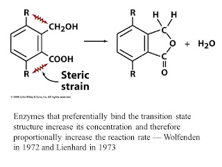
An enzyme may bind to the transition state of the reaction it catalyzes with greater affinity than its substrates or products. the original concept of transition state binding proposed that enzymes mechanically strain their substrate towards the transition states geometry through binding sites into which undistorted substrates did not properly fit. Enzymes that preferentially bind the transition state structure to increase its concentration and therefore proportionally increase the reaction rate.


It is commonly observed that an enzyme binds poor substrates which have low reaction rates, as well as or even better than good ones which have high reaction rates. thus, a good substrate doe not necessarily binds to its enzyme with high affinity, but it does so on activation to the transition state.

Thursday, January 24, 2019

CULTIVATION OF ANAEROBIC BACTERIA:


Stringent anaerobes can be grown only by taking special precautions to exclude all atmospheric O2 from the medium.

1.  Prereduced media: During preparation, the culture medium is boiled for several minutes to drive off most of the dissolved O2. A reducing agent (e.g.,0.05% cysteine, 0.1% thioglycolate, 0.1% ascorbic acid ) is added further to lower the O2 content. Oxygen- free N2 is bubbled through the medium to keep it anaerobic. The medium is then displaced into tubes which are being flushed with O2 free N2, stoppered tightly, and sterilized by autoclaving. Such tubes can be stored for many months. During inoculation, the tubes are continuously flushed with oxygen free CO2 by means of a cannula (inoculum is also added), restoppered , and incubated. This method is also called 'roll tube method’.

2.  Anaerobic chamber: This refers to a plastic anaerobic glove box.


·     Glove parts and rubber gloves allow the operator to perform manipulation within the chamber.
·     Air lock with inner and outer doors. Media are placed within the air lock with the inner door remaining sealed; air is removed by a vacuum pump connection and replaced with N2 through the another pipe.
·     The inner door is opened and the media are placed within the main chamber, which contains an atmosphere of H2 + CO2 + N2.
·     A circulator circulates the gas atmosphere through pellets of palladium catalyst, causing any residual O2 in the media to be used up by reaction with H2.
·     After media have become completely anaerobic they can be inoculated and placed in an incubator located within the chamber.

3. Gas Pack Jar (Anaerobic Jar):
Non stringent anaerobes can be cultured within an anaerobic jar. Media are inoculated and then placed in the jar. Water is added to the gaspack generator envelope causing the evolution of H2 and CO2. The H2 reacts with O2 on the surface of the palladium catalyst, forming water and establishing anaerobic conditions. The CO2 aids the growth of fastidious anaerobes which sometimes fails to grow, or grow only poorly, in its absence. An anaerobic indicator strip (a pad saturated with methylene blue solution) changes from blue to colorless in the absence of O2.


MAINTENANCE AND STOCKING OF PURE CULTURES:

A large collection of strains, frequently referred to as a stock culture collection. These organisms are needed for laboratory classes and research work, as test agents for particular procedures or as reference strains for taxonomic studies. The strains are used for screening of new, potentially effective chemotherapeutic agents; as assay tools for vitamins and amino acids; as agents for the production of vaccines. To maintain strains alive and uncontaminated and to prevent any change in their characteristics, following are the methods for maintenance:

1.  Strains can be maintained by periodically preparing a fresh stock culture from the previous stock culture. The temperature and type of medium chosen should support a slow rather than a rapid rate of growth so that the time interval between transfers can be as long as possible. The transfer method has the disadvantage of failing to prevent changes in the characteristics of a strain due to the development of variants and mutants.
2.  By overlaying cultures with mineral oil: Many bacteria can be preserved by covering the growth on an agar slant with sterile mineral oil. The oil must cover the slant completely; to ensure this, the oil should be about half in above the tip of the slanted surface. This method of maintenance has the unique advantage that you can remove some of the growth under the oil with a transfer Needle, inoculate a fresh medium, and still preserve the original culture. But changes in the characteristics of a strain can still occur.
3.  By lyophilization (freeze drying) : Most bacteria die if cultures are allowed to become dry, although spore- and cyst- formers remain viable for many years. However, freeze drying can satisfactorily preserve many kinds of bacteria that would be killed by ordinary drying. In this process, a dense cell suspension is placed in small vials and frozen at -60 to -78 C. The vials are then connected to a high vacuum line. The ice present in the frozen suspension sublimes under the vacuum, i.e., evaporates without first going into a liquid water phase. This results in dehydration of the bacteria with a minimum of damage to delicate cell structures. The vials are then sealed off under a vacuum and stored in a refrigerator.

Advantages: (1) Many species of bacteria preserved by this method have remained viable and unchanged in their characteristics for more than 30 years.
(2)  Only minimal storage is required; hundreds of lyophilized cultures can be stored in a small area.
(3) The small vials can be sent conveniently through the mail to other microbiology labs when packaged in special sealed mailing containers.
(4) Lyophilized cultures are revived by opening the vials, adding liquid medium and transferring the rehydrated culture to a suitable growth medium.

4. Ultra freezing (storage at low temperature):
In this process, cells are prepared as a dense suspension in a medium containing a cryoprotective agent such as glycerol or dimethyl sulfoxide (DMSO), which prevents cell damage due to ice crystal formation during the subsequent steps. The cell suspension is sealed into small vials are then stored in a liquid nitrogen refrigerator either by immersion in the liquid N2 (-196 C) or by storage in the gas phase above the liquid N2 (-150 C).
Advantage: (i) Many species which cannot be preserved by lyophilization can be preserved by this method.
(ii) Most species can remain viable under these conditions for 10 to 30 years without undergoing change in their characteristics.
Disadvantage: This method is relatively expensive, since the liquid N2 in the refrigerators must be replenished at regular intervals to replace the loss due to evaporation.


METHODS OF ISOLATING PURE CULTURES:

Streak- Plate technique: By means of a transfer loop, a portion of the mixed culture is placed on the surface of an agar medium and streaked across the surface. This manipulation “thins out” the bacteria on the agar surface so that some individual bacteria are separated from each other. When streaking is properly performed, the bacterial cells will be sufficiently far apart in some areas of the plate to ensure that the colony developing from one cell will not merge with that growing from another.

The pour plate and spread plate technique:  In both of these methods, the mixed culture is first diluted to provide only a few cells per milliliter before being used to inoculate media. Since the number of bacteria in the specimen is not known beforehand, a series of dilutions is made so that at least one of the dilutions will contain a suitably sparse concentration of cells.
In pour plate method, the mixed culture is diluted directly in tubes of liquid (cooled) agar medium. The medium is maintained in a liquid state of temperature of 45 C to allow through distribution of the inoculum. The inoculated medium is dispensed into Petri dishes, allowed to solidify and then incubated.
Disadvantages: (a) Some of the organisms are trapped beneath the surface of the medium when it gels, and therefore both surface and subsurface colonies develop.
(b) The organisms being isolated must be able to withstand temporary exposure to the 45 C temperature of the liquid agar medium. Hence, it would be unsuitable for isolating psychrophilic bacteria.

In the spread plate method, the mixed culture is not diluted in the culture medium; instead it is diluted in a series of tubes containing a sterile liquid usually water or physiological saline. A sample is removed from each tube, placed onto the surface of an agar plate, and spread evenly over the surface by means of a sterile, bent glass rod. On at least one plate of the series the bacteria will be in numbers sufficiently low as to allow the development of well separated colonies. In contrast, to the pour plate technique, only surface colonies develop; moreover the organisms are not required to withstand the temperature of liquid agar. Unlike the streak plate technique, the pour plate and the spread plate technique may be performed in a quantitative manner to determine the number of bacteria present in a specimen.


Tuesday, January 22, 2019

MICROBIAL PRODUCTION OF LIPASE:


lipase catalyses the hydrolysis of dietary fats like triglycerides into fatty acids and glycerol.

Source: lipase is produced by bacteria like Pseudomonas aexuginosa as well as by fungi like Aspergillus flavus, A.niger etc.

Types of fermentation: solid state fermentation and submerged fermentation. However, solid state fermentation is frequently used.

Production parameters:
A. Pseudomonas aeruginosa

Composition of production medium:

Component.                     Percent
Yeast extract.                     0.3
Peptone.                            0.1
Olive oil.                              1
K2HPO4.                          0.07
KH2PO4.                          0.03
MgSO4.                            0.05
MnCl2.                             0.01
(NH4)2SO4.                    0.025
CaCl2.                               0.01

Optimum pH for production is 7.2 using 0.5N NaOH.
Optimum temperature is 35 C with constant shaking at 125 rpm.
Fermentation period: 72 hours.

Downstream processing:
Culture medium is centrifuged at 10,000 rpm at 4C for 20 minutes.
Centrifugation is followed by salt precipitation method
Purification is done by column chromatography method.

(B) For fungal sources:

Table

Downstream processing:
To extract this enzyme, substrate is homogenized or mixed in a rotary shaker with one of the solvents including water, NaCl, (NH4)2SO4 and NaCl with Triton X-100.
The mixture is then filtered using a muslin cloth and the filtrate is centrifuged at 5000 rpm for 20 minutes around 4 C.
Above steps are followed by concentration of the filtrate using salt precipitation and finally purification of the enzyme by column chromatography.

APPLICATIONS:
In the detergent industry, lipases remove oil stain from fabrics by hydrolysis of fats.
In the food industry, lipases play an important role in flavor improvement, aroma quality improvement. Transestrification, fat removal, shelf- life prolongation.
In the paper and pulp industry, lipases improve the paper quality and improve pulp strengthening.
In the leather industry, lipases are involved in softening, quality improvement in the process of tanning.
In the chemical Industry, lipases play an important role in transestrification, hydrolysis, synthesis, enantioselectivity and thus involved in cosmetics as well as digestive aids production.
In medical field, lipases can be used to diagnose acute pancreatitis. Lipase can assist in fat breakdown into lipids in those undergoing pancreatic enzyme replacement therapy.

PHASE CONTRAST MICROSCOPY

Introduction: Most cells are too small to be seen by the naked eyes, the study of cells has depended heavily on the use of microscopes. Mi...